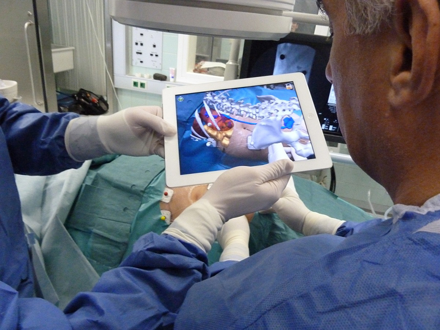
In order to help surgeons perform image-guided open and minimal-invasive spinal surgery, Royal Philips works on an surgical navigation technology based on augmented-reality.
According to a company statement, the technology is significant for cranial and trauma surgeries, pediatric spine surgery, and thoracic spine fusion surgery in adults. Because the thoracic spine is located in the mid- to upper part of the back, the vertebrae are smaller than that of the lower lumbar spine. This makes thoracic fusion surgery more difficult and risky for the surrounding nerves and tissue.
In a preclinical study, more accuracy in placing screws in spines of cadavers, was obtained by neurosurgeons using augmented reality. The accuracy rate of pedicle screw placement in thoracic cadavers’ spines versus freehand placement was increased from 64% to 85% according to the study result.
Less complications are implied by better accuracy according to the business leader of image-guided therapy systems at the company located in Amsterdam.
Since the release of Google Glass in 2013, surgical applications for augmented and virtual reality have gained attention. A London surgeon and the co-inventor of a VR company used the low-cost VR system Google Cardboard to livestream a procedure removing a colon tumor from a patient in April 2016.
In order to add its latest technology, for clinical trials scheduled at about 10 global testing sites, together with its existing low-dose X-ray systems, Philips will need regulatory approval. After full approval by FDA and EU, Philips can expand its use to countless hospitals around the world.
To image the surface of the patient a high-resolution optical camera is placed on a flat X-ray device. The technology constructs a 3D augmented reality vision of the internal and external anatomy, by combining the internal 3D X-ray view and the external camera view. This actual 3D image of the spine of the patient improves procedure planning, surgical tool navigation, accuracy of the implant, and reduces procedure time.
Especially in cranial surgery the technology could be useful, because of brain shape change after releasing the pressure in opening of the cranium. This makes actual images more important than pre-surgical MRI or CT scan images.
Because of the extensive experience in the field of designing image guiding systems Philips needs to tailor them to specific procedures. Starting with spinal surgery, fine tuning the system for trauma surgery will follow, and in the future there is the possibility of the system assisting in brain surgery.